(Originally posted October 31, 2019, then October 31, 2023)
Date for this post: Oct 1, 2024
Mammography schedules are jammed in October. It’s hard to work in the callbacks and biopsies. Everyone is pressured to get it done before the looming Holiday Season. And if we find cancer, it spoils that festive stretch from Thanksgiving to Christmas, Hanukkah, or whatever, through New Years…not to mention the fact that deductibles were met at the time of the mammographic screening, but then in January, it starts over again as cancer care continues. Ugh! How did we ever get here?
Prior to 1980, there were no pink ribbons, no races, no walks. There was very little awareness and breast cancer was not discussed in polite society. The fact that Shirley Temple Black (breast cancer in 1972), Betty Ford (1974) and Happy Rockefeller (diagnosed 2 weeks after Ford in 1974) made their diagnoses public was shocking (and trailblazing). And when the Susan Komen Foundation was established in 1982, newspapers resisted using the words “breast cancer.” As late as 1993 when we opened our doors at the University of Oklahoma Institute for Breast Health, there were complaints about our signage with its “irreverent” word BREAST plastered right there in public.
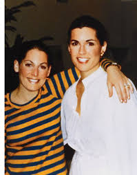
Let’s go back to the 1940s when Susan and Nancy Goodman were sisters growing up in a well-to-do Jewish family in Peoria, Illinois. Who could have conceived of the notion that both girls carried a mutation in BRCA-1 that would lead to breast cancer in the both of them? After all, it would be more than 50 years before the launch of commercial testing of BRCA-1 and BRCA-2.
(Susan Komen on the left; sister Nancy Brinker on the right)
Susan grew up to be the “darling of Peoria,” a beauty queen and local model. She would enter a disastrous first marriage (groom collapsed at the wedding for starters), followed by a second marriage to Stan Komen who would run a liquor store – Stan’s Wine and Spirits – in Peoria until his retirement in 2014.
At age 33, Susan felt a breast lump. Not a good thing in 1977, on the eve of a revolution about to occur in the management of breast cancer. 1977 was, however, the peak time of enthusiasm for “subcutaneous mastectomies” with the newfangled breast implants for reconstruction…sometimes patient-driven, sometimes surgeon-driven and sometimes both. Pain, cysts, “fibrocystic disease,” you name it, then cough up the money, and surgeons would perform the “scoop out” procedure with implant reconstruction. Some women were very happy with their result. Many were not. And for some, considerable amounts of breast tissue remained in place. Nevertheless, no one at the time conceived of using the procedure for treating cancer — except for Susan’s surgeon who had been recommended by her family physician in Peoria.
After performing subcutaneous mastectomy for Susan’s cancer, the surgeon pronounced her cancer-free, a tad premature since she developed positive nodes shortly thereafter and systemic metastases as well. After treatment at both the Mayo Clinic and M. D. Anderson, she was still left with chest wall recurrences and died 3 years later in 1980 at the age of 36. Given the aggressiveness of this tumor, it is hard to lay blame on the unusual subcutaneous approach used for local control, but it is noteworthy nonetheless.
Meanwhile, younger sister Nancy had moved to Dallas where she became a buyer for Neiman Marcus, and then, in 1981, she became the bride of Norman Brinker, the restaurant magnate who founded Steak and Ale, et al, (now Brinker International) and is credited with the development of that omnipresent dining staple – the salad bar. With her new financial stature and a deathbed promise to her sister Susan, Nancy Brinker founded the Susan G. Komen Breast Cancer Foundation in 1982, a mere 2 years after Susan’s death. The foundation became intimately linked to Dallas as a result of Nancy’s new home, not Susan’s home in Peoria (although Peoria did become a site for a “Komen Breast Center,” a nationwide network concept of screening centers that never happened.)
The first Race for the Cure took place in Dallas, Texas with 800 participants in 1983. The rest is history. I attended the first race in Oklahoma City (1989, as I recall) where participants raced around the track at Remington Park. Pink ribbons were not introduced until 1991, and though several groups lay claim to the pink revolution, I’m pretty sure it was a Komen innovation.
One year after that first Race for the Cure, Nancy Brinker was diagnosed with breast cancer. She underwent unilateral mastectomy and later contralateral prevention, even though her BRCA-1 mutation was not confirmed until 2006.
Initially, the sole agenda for Komen was to flood the country with high quality screening mammography. This drew some objections, including one prominent breast surgeon, the late Susan Love, who broke ranks with the movement by clarifying that mammography is not a “cure,” and that we needed to be thinking more about a true cure and a “post-mammographic era.” Eventually, Komen expanded its scope to all types of breast cancer research.
Nevertheless, the growth of the Race for the Cure was phenomenal, taking place in a parallel fashion to the breast cancer diagnosis and treatment revolution that was well underway. It is estimated that, today, over 1.5 million participants raise money through one of several outreach programs sponsored by Susan G. Komen (several name changes of the foundation have occurred over the years, but Susan’s name is always there). Politics, of course, wormed its way into the activities of Susan G. Komen and eventually Nancy Brinker resigned as CEO.
Back to Breast Cancer Awareness in the spooky month of October. Around 1985 or 1986, the American Cancer Society teamed up with a pharmaceutical company that later became Zeneca, then AstraZeneca, announcing October as National Breast Cancer Awareness month. Zeneca was criticized for self-serving interest since they manufactured pharmaceuticals used for breast cancer. But they had, in fact, done their own internal audit on employees, showing that it was cheaper to screen with mammograms for an early diagnosis than to do nothing and pay for treatment of more advanced disease.
At this same time, the famous (or infamous) Breast Cancer Detection Demonstration Project was reporting results indicating that massive screening of the general population in the U.S. was both feasible and effective. The BCDDP was sponsored by the American Cancer Society and the National Cancer Institute, both organizations riding the waves of the War on Cancer legislation signed by President Nixon in 1971. So, by the mid-80s, the policy makers were wildly enthusiastic about general population screening with mammography, and it was a case of “full steam ahead.”
Still…why October?
I’ve not been able to nail down the exact details surrounding October as Breast Cancer Awareness Month, but I have a theory — a link between the rapid rise of the Susan G. Komen Foundation and the proclamation about October made by the American Cancer Society. I think the answer might be found by looking at Susan Goodman Komen’s birthday – she was born on October 31, 1943. She would have been 81 this Halloween.
In 1976, pioneering breast radiologist, John Wolfe, published results he’d been working on for the prior decade — that is, certain mammographic patterns being associated with an increased risk for breast cancer. This wasn’t your measly “4-6X” risk that emerged (with much confusion) in modern times, but a 37-fold increased risk imparted by the now-infamous “DY” pattern. Translated to absolute risk, that’s 45% breast cancer risk for women aged 40-59, and a whopping 82% risk if focused only on the 50-59 age group. And with that in mind, Dr. Wolfe recommended that those women identified with the DY pattern undergo preventive subcutaneous mastectomies, a new procedure being promoted by the plastic surgeons (for a variety of indications) upon the introduction of silicone implants.
The curious thing about Wolfe’s landmark article and his recommendations is that he divided mammographic patterns into 4 categories (the same number we have today), but the divisions were based on breast cancer risk, NOT the likelihood of missed cancer, i.e., “sensitivity.” The N1 category was “predominantly fatty” and was associated with such a low cancer risk that screening was not needed. P1 meant less than 25% “prominent ducts” and again, low risk for cancer, so no pressing reason to screen. P2 was defined as greater than 25% “prominent ducts” and screening was now advised due to the risk status. DY was dense fibroglandular tissue with very high risk (analogous to today’s Level D), to the point that Wolfe made his remarkable recommendation for preventive surgeries for all DYs. In 1980, when I began private practice in Los Angeles, all mammography reports included the Wolfe classification, though we had no idea what purpose it served since interventions were unproven, and screening recommendations were new. None of the recommendations from various organizations included Wolfe in their guidelines.
Turns out, Wolfe’s methodology was flawed for a variety of reasons. First of all, most of the women he studied had symptoms of some sort. That is, they were not asymptomatic, which is required today before calling a study “screening.” So, for example, when a patient in his study with a DY pattern developed breast cancer 7 months after a negative mammogram, Wolfe felt the DY pattern, as a strong risk factor, had accurately predicted a future cancer. But this wasn’t the case at all! He was witnessing the extraordinarily high “miss rate” for mammography when women have dense breast tissue. The cancer was there on the prior mammogram, but not visible due to the camouflaging effect of density! He was correctly observing a relationship between density levels and breast cancer, but misunderstanding the true reason for his observations. Ever since then, we have struggled to recognize that there are two related, albeit independent, implications of high mammographic density — 1) a modest risk factor, and 2) the strongest predictor for missing a cancer on routine screening (“poor sensitivity”).”Wolfe patterns” came and went, and after that, trying to relate breast cancer risk to mammographic patterns fell into a state of hibernation. Any attempts to document increased risk associated with density patterns went unnoticed by clinicians. We had moved “beyond Wolfe,” it seemed. At the same time, an enormous effort was engineered by the American Cancer Society and others to promote mammographic screening. In these efforts, mammographic sensitivity was said to be “90-95%.” As it turns out, this “90-95%” was based on a large feasibility study (the BCDDP, not the randomized controlled trials that proved screening saves lives) where many of those entering the massive BCDDP study had palpable masses. Stage II cancers outnumbered Stage 0 and Stage I combined. “90-95%” was never true for asymptomatic women, yet it dominated thought for another decade or so. Few clinicians were paying attention to the rather large number of cancers that were being missed on screening mammograms. This should be no surprise. How do you recognize and count the number of missed cancers if you’ve missed them already? The standard approach was to count cancers that emerged after negative mammograms for a period of 12 months of follow-up, but this was arbitrary and unreliable. (Adjunct imaging to identify missed cancers was not yet available.) For palpable cancers — yes, the sensitivity was 90%. But that’s not screening, and we failed to make that distinction in those early years. The definition of mammographic screening evolved to imply asymptomatic patients (no palpable masses), and it was here that the sensitivity of mammography was largely unknown to clinicians.
Then, technical developments took place in the 1990s with ultrasound (US). No longer was US just a matter of cyst vs. solid, but it could distinguish benign vs. malignant. And with this development, many radiologists launched studies to screen asymptomatic women with US, after negative mammograms…IF the breast tissue was dense. There were no definitive trials at first, that is, no prospective randomized studies. But the results with US were numerically consistent across the board. Mammograms weren’t coming close to 90-95% sensitivity in women with dense breasts. Completely independent of “future risk,” breast density was predicting mammographic failure due to the camouflage effect. Arguably, this failure to detect cancer is much more significant than the modest risk imparted by density (more on this below).
Then, in the early 2000s, the introduction of MRI into the breast clinic turned the sensitivity problem upside down — that is, mammography alone in high-risk women was missing more cancers than it was finding. Sensitivity was 40% across the board in the MRI screening trials. This was a far cry from the “90-95%” starting point 25-30 years earlier. Granted, MRI lowered the threshold of detection to smaller tumors, which made both mammography and US look worse. On the other hand, ultrasound did not lower the threshold of detection, so for purists, US probably reflects the miss rate of mammography more fairly. Nevertheless, what MRI is doing becomes clear when the numbers are examined — MRI is finding current breast cancers that are invisible on mammography and/or US, then — by lowering the threshold of detection, it is also finding most of the breast cancers that won’t be discovered until the next routine screening study, 1 or 2 years away.
Back to Breast Density. Overcoming the “false alarm” by Wolfe was not easy. I was once part of a team of primarily academic radiologists who taught MRI to other radiologists around the country, shortly after MRI was introduced. I was the token surgeon. In one of my talks on MRI screening guidelines, I pointed out the overwhelming evidence that breast density was an independent risk factor for breast cancer development. This was nowhere near the Wolfe levels, but the positive results were similar in more than 10 studies. So, why was no one paying attention? I don’t know. Were they remembering the collapse of the Wolfe criteria? In my presentations, I likened breast density to comedian Rodney Dangerfield — “No Respect.” Breast density was not part of the mathematical models that estimate risk. It was not used clinically with regard to any screening recommendations with any modality (It’s still not considered a problem requiring multi-modality imaging by the U.S. Preventive Services Task Force!). And, when added to the MAJOR problem of failing to detect breast cancer, density was — well — it was Rodney Dangerfield’s calling card: “I don’t get no respect.”
I don’t think I made a dent in the lack of interest in breast density. What made the difference was a grassroots movement launched when women discovered the truth – that many of them had mammograms so dense that cancer detection was nearly impossible, yet no one told them! (I’ve covered this story about the late Nancy Cappello in other blogatorials.) Today, we celebrate legislation that forces radiologists to talk to patients about this problem, but how in the world did we get to the point where this had to be legislated? I started a High Density counseling program and clinic at my facility just as soon as we opened our MRI screening program in 2003. And, I published a point system for MRI screening where Density played an equal role to Risk level. In my mind, density was even more important than risk level. After all, why talk about a second form of imaging if you don’t take into account the failure rate of the first modality?
Yet, when the American Cancer Society released their 2007 guidelines for MRI screening, “Density” was relegated to the “need more research” category. And it’s been a brick wall ever since. We also published our local MRI screening results before and after the ACS guidelines, wherein one-half of our MRI-discovered cancers would not have been found if we’d followed the ACS guidelines. By including density, our MRI cancer yield doubled. Europe is way ahead of the U.S. on this one. The European Society of Breast Imaging has recently added a screening approach for women with Level D density, with no other risk factors: “Breast MRI every 2-4 years.”
So, even though radiologists must divulge high density levels to patients, not everyone will counsel patients at that point as to options. Even if options are discussed, the breast center must understand that insurance often will not pay for high-density screening beyond mammography (following the Task Force guidelines). So, the breast center should have low-cost programs for cash customers who have opted for US or MRI (but that’s another story — many hospital-based breast centers don’t like that “low-cost option.”)
Now, in spite of the powerful presence of “breast density” in the screening world today, there are points of confusion still circulating. I’ll mention 6 of them:
“Density imparts a 4-6 fold risk of breast cancer.” This is highly misleading, as this is a “relative risk,” and all relative risks have a numerator and a denominator. In this case, the denominator is those women with fatty replacement pattern (Level A). This is only 10% of the population and hardly “average.” So, yes, comparing Level D to Level A generates “4-6 fold risk.” But if you compare Level D to the “average patient” then the Relative Risk becomes a much more manageable 2-fold risk. This misconception was so pervasive initially that huge efforts were made to educate physicians about these numbers, and the problem has been largely corrected. Most clinicians have been exposed now to this misconception, though it still leaks out occasionally.
“The risk imparted by density is now covered nicely by the Tyrer-Cuzick (TC) model.” Careful. The referent (where RR=1.0) in the TC model is essentially Level C density. Not Level A. Not Level B. So now, patients who have been labeled as “dense” Level C and a slight increase in breast cancer risk will actually have Zero risk (RR=1.0) added by density using Version 8.0 of the TC model. So, “high risk” when speaking of dense breasts in general, but “no elevated risk” for Level C when using the TC model. Only Level D translates to higher absolute risk in the TC model. Level C is neutral, while Levels A & B will actually generate lower levels of risk with the newer Version 8.0 (RRs less than 1.0).
“Dense and non-dense is the currently preferred approachin defining women for specialized screening.” What we really have is a density continuum from 0 to 100% (the practice of medicine “hates” continuums, as we need to stratify our strategies.) Furthermore, we have a classic Bell Curve, with most patients bunched up in the middle where the Level B and Level C patients dominate, accounting for 80% of the total. So, when we turn this into a dichotomy, we draw a line down the middle of the Bell Curve, and there is little difference between a high Level B and a low Level C. In fact, it is highly subjective, and radiologists don’t necessarily agree; even a single radiologist can be inconsistent. Yet, this is where most women are due to the Bell curve — either “high B or low C.” But the “dense and non-dense” dichotomy (A/B or C/D) implies a sharp distinction that’s just not there.
“Software is now available to take away the subjectivity.” While it is impressive to see exact percentages emerge with these tools, you can’t get away from the Bell Curve. Most women are clustered right at the division line, with huge differences in management depending on which side one falls. This limitation (confusion?) has been created through the simultaneous use of a dichotomy (by the grassroots movement) and the 4 Levels A-D from the American College of Radiology. If we’re going to use 4 levels, then each level should have unique recommendations. Another problem that is not addressed by software packages — there is a qualitative feature of density in addition to the well-known quantitative. This qualitative aspect is exceedingly complex and beyond discussion here. However, to give a practical example, I create a dime-sized mass in my head, then move it throughout the white areas of a mammogram to see if there’s somewhere a cancer could hide, even in low density patients. If you are a clinician, think about those mammograms where the only density is a homogeneous white sheet confined to the upper outer quadrants. This will be a Level B patient with only 25% density, but if cancer develops in the UOQ, it’s going to hidden, just as if the patient were Level D. Remember, overall breast density is merely a surrogate for the density that surrounds a cancer. And, this could even be a worse problem, considering that these dense patches might be where cancer originates. (P.S. If you think this sounds overly compulsive (nuts), then read about the Entry requirements for ACRIN 6666 (density + one additional risk factor, studying multimodality screening). A patient could qualify with a low overall density if there was a homogeneous white region involving just one quadrant in each breast. Level A in 3 quadrants, Level D in one quadrant, but instead of a fusion to Level B, patients qualified for ACRIN 6666). You can see that the risk of missing a cancer on mammography has this qualitative aspect in addition to the 4 quantified levels.
“Density disappears with Age.” Radiologists know this isn’t true, but many clinicians (and epidemiologists who set screening guidelines) believe that extra imaging with US or MRI will not be needed after a certain age (60, for example) because low density translates to mammographic accuracy. While this transition to less density might be true overall in a large cohort, there are many exceptions to this rule. We’ve all seen the 70 y/o with Level D density. And, when encountering the young woman with Level D density, it might improve somewhat over time, but she is going to remain dense her whole life, even though a shift down is possible. This controversy comes up in the MRI screening guidelines where older women are discriminated against with “lifetime risk” calculations. Older women have passed through much of their lifetime risk, so they don’t qualify for MRI screening, yet their short-term risk might be very high — much higher than a younger woman who qualifies on the basis of many years left in her lifetime risk. I’ve tried to make this point in publications and at screening guideline meetings, and am met with, “the MRI guidelines were intentionally designed for younger women with dense breasts.” Fine. Agreed. But what about the older woman with dense breasts? They can be included without taking away any of the benefit for younger women. And, the answer is so easy — just add short-term risk calculations alongside the lifetime calculations. On this point, I’ve made about as much headway as Rodney Dangerfield.
“3D tomosynthesis has solved the density problem.” Or, the way a patient might state the issue — I’d like to stop Ultrasound (or MRI) now that we have 3D mammography.” Sorry, but 3D is not even close to solving the density problem. It helps, yes, but let me put this in perspective. Shortly after the introduction of 3D, an Italian study pitted 3D mammography compared to old-fashioned 2D plus Ultrasound. It wasn’t even close. The combination of 2D plus US discovered many more cancers than 3D. It gets worse. A clinical trial in the U.S. pitted breast MRI against 3D mammography. MRI found 95.7% of the cancers while 3D found…wait for it….39.1%.
Is there any hope for the patient with dense breasts, short of MRI? Yes. While Ultrasound is currently thought to be the next best thing to MRI in high-density screening, Contrast-enhanced mammography (CEM) is more sensitive than ultrasound and might be the answer. We’ve already seen the numbers. CEM is using a radiologic dye, injected prior to the 3D mammogram. Results appear to be in the same ballpark with MRI, though more definitive studies are being performed now. We know enough already that CEM can be recommended when patients are unable to undergo MRI for some reason. And, CEM is easier on patients, and cheaper. The question is whether or not it can replace MRI rather than its current role as a comparable back-up.
PS – I’d be remiss if I didn’t mention the ultimate answer (in my mind) to all the mess above, and even more mess I haven’t addressed with using Risks and Density to guide adjunct imaging recommendations. If we had a reliable screening blood test, then women could undergo mammography and blood testing as a matter of routine. And if mammograms were negative, but blood test positive, this would be the signal for adjunct US, CEM, or MRI. (That’s why I’ve been pursuing this agenda for the past 30 years.)
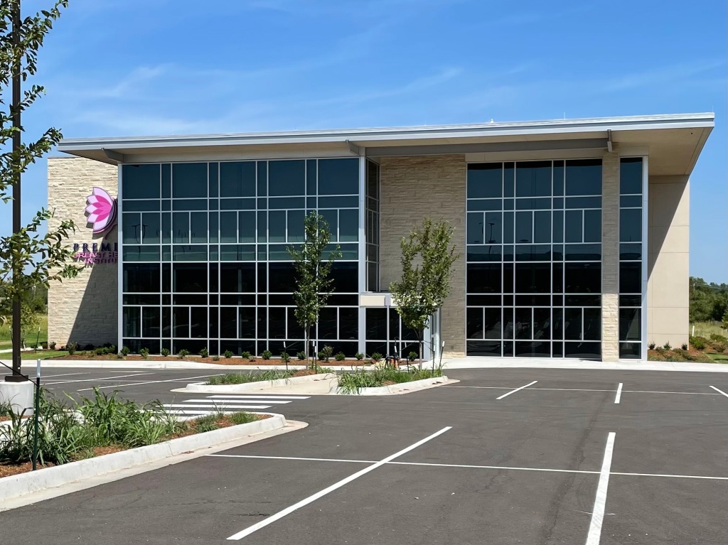
I’m going to end it there, but with a plug for a new breast center opening soon in OKC (SW 89th & I-44), where we will have Contrast Enhanced Mammography as an option, as well as a High-Density clinic to accompany the High-Risk Clinic. More information to follow, but the Premier Breast Health Institute of Oklahoma will be opening its doors in the fall, 2023.