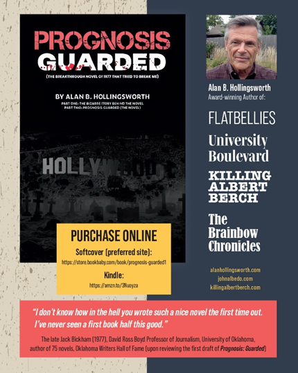
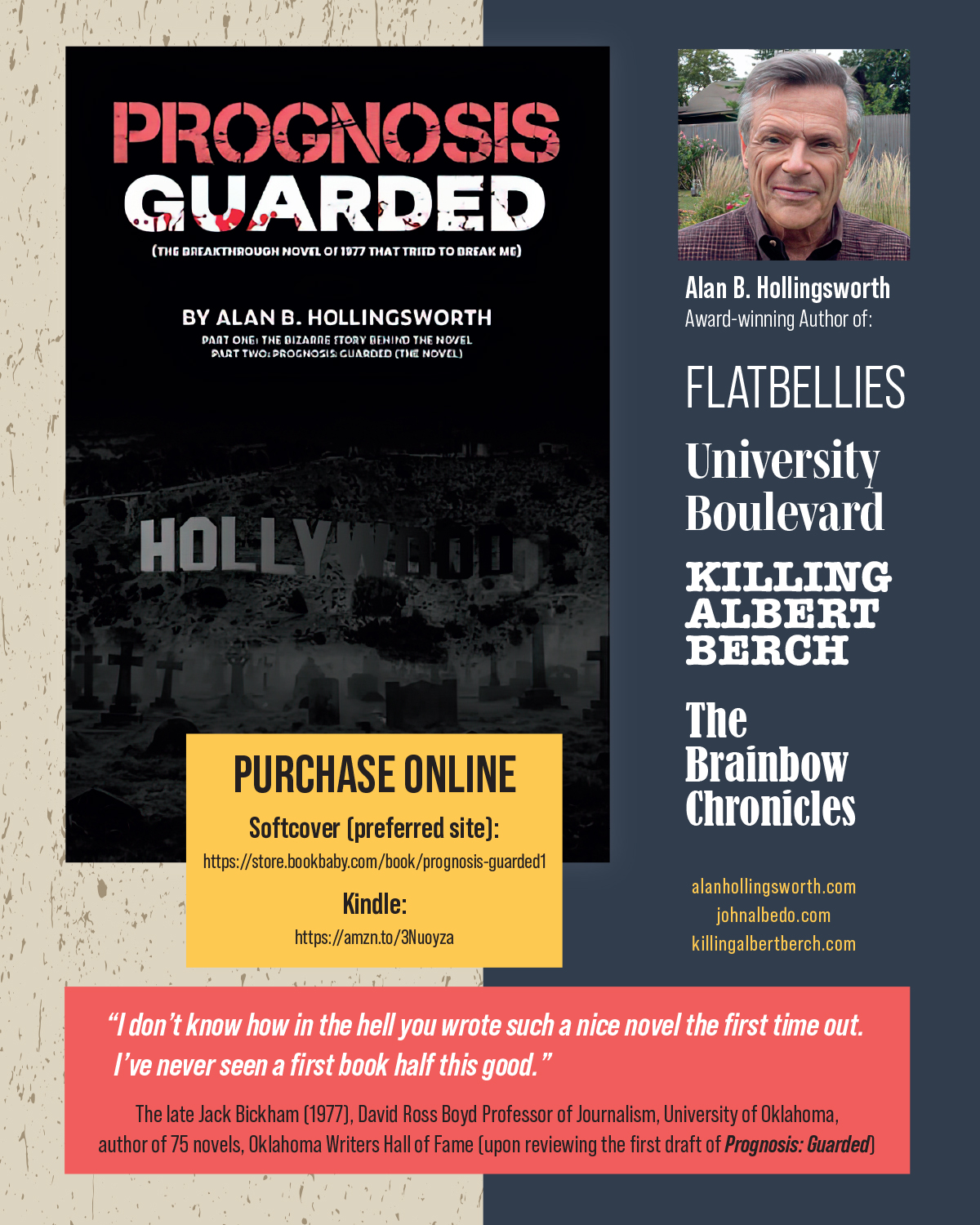
Is PROGNOSIS: GUARDED (PG) a prequel to FLATBELLIES?
No. Although PG was written much earlier (first draft 1977) than FLATBELLIES, the story in PG is unrelated to FLATBELLIES, published in 2001. That said, Part One in this “new” PG is the true story of a 17-year effort to get PG published, to no avail. Ordinarily, such an account would be ho-hum, as literary rejection is far more common than literary acceptance. But there’s a difference here — the original PG was pegged as a literary blockbuster “soon to be a major motion picture.” The story of what happened after that first draft in 1977 was a life-changing experience for the 29 y/o author (me). And, the collapse of the original PG led to a major switch in genres on my part, dropping the medical thriller plans and adopting coming-of-age fiction. Thus, Part One of the “new” PG is an “origin story,” explaining the emergence of FLATBELLIES. Part Two is the medical thriller that caused all the controversy and confusion, in print for the first time.
What is the book’s cover trying to show?
The famous Hollywood sign is disappearing, and at the base of the sign is a cemetery. The cover is representing both Part One and Part Two. Part One of PG is the death of a Hollywood dream. Part Two of PG is the novel that deals with orchestrated medical murders of the rich and famous of Hollywood. (In some of the printings, the cemetery at the base of the Hollywood sign is dark and hard to see.)
Is the story of PROGNOSIS: GUARDED pure fiction?
Yes. It’s the only one of my six novels that did not borrow from actual events.
Why isn’t PROGNOSIS: GUARDED available at my local bookstore?
Long story, but I felt that combining Part One and Part Two into a single book (one non-fiction and the other fiction) was going to make traditional publishers squeamish. So, I went the route of publishing through an Amazon resource that gave me full editorial control, but also requires sales to come through Amazon. Depending on how it goes, I might make arrangements with booksellers in OKC to sell PG, as we are ramping up for a 25th anniversary edition of FLATBELLIES, and PG is the start of that process by telling the origin story first. So, if we release the special edition of FLATBELLIES next year, it would likely be sold side by side with PG at bookstores.
Kindle purchase per Amazon, or however you are accustomed to buying ebooks.
Do you still speak at book clubs?
Yes. I attend any book club within a 2-3 hour driving radius of OKC. Anything beyond that, I do online. Also, some clubs are not specifically book clubs (social clubs, study clubs, etc.), yet they will ask me to speak about writing and publishing in general, or to discuss one of my novels from the past (see http://www.johnalbedo.com). Or, there is great interest in my one true crime story covering the 1923 murder of my grandfather (see http://www.killingalbertberch.com). Contact me through Facebook Messaging to schedule. Or, email alanhollingsworth@cox.net.
I read COMA when it first came out, and now that I’ve read PROGNOSIS: GUARDED, I don’t think the two books are that much alike. What gives?
Recall from Part One that I don’t have a copy of the original 1977 manuscript, which was virtually identical to COMA, complete with suspended human bodies. Several New York publishers (most notably St. Martins Press) asked me to change my version as much as possible to allow publication as an original story. The 1980-81 version I found in the attic (the published version) was the end product of that effort, taking away the science fiction component and transferring the genre to a medical murder mystery. I thought the changes were adequate, but publishers still ruled it “too much like COMA.” This was likely due to the fact that the “medical thriller” was still a new concept, so my revised version was compared to the only other successful thriller at the time — COMA. Had there been 100 medical thrillers on the market, I think PG would have had no trouble falling into that pool. (Remember this from Part One — when Random House rejected my book as “too much like COMA,” the Senior Editor called PROGNOSIS: GUARDED a “hospital intrigue story.”)
NOTE: December 18th is the official launch by Amazon. Softcover can be ordered from the same page as Kindle (above), but delivery won’t be until after December 18.
My “new” book release is PROGNOSIS: GUARDED, a medical thriller drafted originally in 1977, making the novel nearly a half century old. Of all the author duties that accompany a book launch, the worst is “begging for stars” on Amazon Customer Reviews. Somehow, the star system (under 4 stars, the book does not exist) has bypassed professional reviews, making the Customer Review the most important parameter other than actual sales. So, if you read the book — available in softcover and Kindle, sold only online (see below) — and feel inspired, here’s the link to Customer Reviews: https://amzn.to/3Nuoyza
To help explain the 47-year gap between the first draft and publication, I added an introductory segment, such that our marketing “blurbs” read something like this:
Terror is timeless. This 1977 medical thriller will have you turning pages faster than you can say “Robin Cook” (whose 1977 novel COMA defined a new genre). But what happened to PROGNOSIS: GUARDED between 1977 and 2024? PART ONE takes the reader on a wild ride through the world of publishing and the life-changing impact the book had upon the author whose original draft was a locked and loaded blockbuster (or so everyone thought). PART TWO is the novel itself — PROGNOSIS: GUARDED, still crazy after all these years.
“Early detection is the key,” we all say. But is it really that straightforward? In the late 1980s, one of the most influential surgeons in the history of breast cancer management (Bernie Fisher, MD) was at the podium defending his theory of breast cancer biology, in support of lumpectomy, when he said: “I don’t know what early breast cancer really is. There’s no satisfactory definition.” Mammography had hit the scene, prompting the term, “mammographically-detected cancers,” but Dr. Fisher was adamant that his theories that justified lumpectomy were not dependent on the method of tumor detection. Furthermore, just because a cancer was identified through screening mammography did not necessarily mean it was “early.”
(For those who believe that screening mammography was the primary reason behind breast conservation, or that pre-op mammography was part of the “package” tested in the landmark NSABP B-06 trial, you might be surprised to know that accrual to those studies started in the 1970s and did not require that mammograms be performed. Lumpectomy arose from a biologic theory that pre-dated mammography, and would have confirmed the safety of lumpectomy with or without mammography.) That said, if Dr. Fisher didn’t know the definition of “early breast cancer,” then who am I to attempt a definition? And, yes, I remember that it’s the umbrella name of this blog.
Bernard Fisher, MD
The problem with the term, Dr. Fisher explained, was that small tumors can still be deadly, and large tumors can be indolent, so using tumor size for the definition was not reliable. And if we try to use a measure of time to represent early vs. late, we are equally lost since we really don’t know how long the tumor has been present. He maintained that the outcome (cure vs. no cure) was based on the inherent biology of the tumor and how it interfaced with the “host’s” immune system. Both tumor and host were equally important. Since he came up with that theory from laboratory studies in the 1950s, it’s hard today to call this concept “new.” Yet, given the immense amount of research now being performed, not necessarily on the cancer cells, but on the immune cells that govern the host reaction, one can say that the late Dr. Fisher was a scientific prophet or, at least, remarkably prescient.
I bring this up because I was recently surprised by a “new” use of the term “early.” In fact, within the context I’m going to give you, “early” was used for what I’ve always called “late,” or even “locally advanced.”
Recently, I attended the 46th annual San Antonio Breast Cancer Symposium, a spectacle that every breast specialist should attend at least once (I think this was my 20th time). Over 10,000 attendees from 102 countries, with literally 1000s of posters and presentations over a 5-day period. The conference has always been skewed heavily toward medical oncology and its associated research, now in high speed with the development of countless drugs that alter the immune system to attack cancer cells. And all power to them. But if you’re looking for new developments in breast imaging, surgery, radiation, plastics, prevention, or basic pathology, this is not the conference for you. These topics are relegated to the poster sessions, rather than the podium. Even more than before, it has become a medical oncology event, along with related research. Admittedly, one reason I attend is to meet with international collaborators who have made the trip to San Antonio. In fact, the first 3-4 times I attended, I went to meetings “on the side” and didn’t even enroll in the course itself.
This time around, I had not attended for three years, so you’d think not much had changed. But it has. There is a shifting tide, a subtle sociologic phenomenon, that I can’t find the right words to describe. It’s easier to point out that we were told: “Given physician stress levels of today,” we attendees would have access to massages, yoga, etc. And, “if you need to up your brand,” there was a professional photographer stationed in the event center who would give you the “head shot” of your choice. The line of scientists and clinicians hoping to “up their brand” was long and was populated by attendees who appeared as teen-agers to me. A quiet voice whispered: “This is no country for old men.”
Back to the conference sessions. One by one, researchers and clinicians approached the podium and announced the title of their presentations, many of which contained the phrase “early breast cancer.” For a generic example, “Thank you for that wonderful introduction. I will be presenting today the final results of the XXX trial that administered YYY in combination with ZZZ versus YYY alone in the pre-surgical neoadjuvant therapy of early breast cancer. Over and over, it was early, early, early, while I’m seeing late, late, late. Through societal forces too complex for my blood, in the past 3 years, it had become a conventional norm to apply the word “early” to any patient with breast cancer who did NOT have known metastatic disease. That is, any patient with Stage I, Stage 2, or Stage 3. It seemed the only time “late” would have been used was if study patients had Stage 4 disease.
So, as the researchers would announce life expectancies extended by 3 months, or 5 months, or 8 months, through the use of the new ZZZ compound, I had to think what would have been the results if every participant in each particular trial had been on a long-term program of breast MRI screening at 1-2 year intervals? In fact, we’re only now seeing the “cure rates” for breast cancer when discovered by MRI, but it appears that very few patients would have qualified for the ZZZ trial because their disease would have been “too early.”
Debate and analysis about the relative benefit of systemic therapy (by medical oncology) vs. screening (by radiologists) has, in the past, called it a tie — that is, both approaches reduce breast cancer specific mortality by the same amount. But that’s mammography, where half the cancers are missed when breast tissue is dense. Screening with MRI is a whole different ballgame, yet it is held in check due to outdated guidelines (that don’t include breast density…yet), and the impracticality and cost of screening the entire population with MRI.
But I digress. My astonishment was in the new definition of “early.” And it speaks to the remarkable confusion imparted by different meanings for the same word or words. Brilliant scientists somehow get comfortable with the ambiguous lexicon used by their particular sub-specialty, while colleagues from different camps are using the same word for something else. Oddly enough, I’ve published an Editorial on this very thing.
In 2015, the Editor-in-Chief of The Breast Journal asked me to write a critique about the lead article, which was going to be published soon. Such an analysis of a lead article is a time-consuming honor, by the way, that counts very little for anything, other than knowing your opinion is respected by at least a few. But when I read the article about to be published (dealing with the implications for surgery and radiation when patients have more than one location of tumor in the breast), I asked if I could use a major deficiency as a springboard for another topic entirely — the power of language (a theme in my novels, too). The authors, all from a prestigious academic center, had used terminology that meant different things to different people. Specifically, no attempt was made to distinguish the varied definitions of “multifocal” vs. “multicentric,” in my view, imparting a great deal of confusion for any reader. Same thing for the term “local recurrence,” which drags around several different definitions as well.
The result was an Editorial titled, “The Beginning of Wisdom is the Definition of Terms,” a phrase I borrowed from Socrates.
Some of my colleagues around the country got a kick out of the tongue-in-cheek editorial and emailed their responses, and that was the end of that. But 2 years later, a notification came through the mystery of the internet, stating that I had been quoted in the Journal of Volcanology and Geothermal Research. Must be a different Alan Hollingsworth, I thought. Nevertheless, I looked it up, and sure enough — I’d made it into the world of volcanos. As it turned out, this article was an appeal for the different sub-specialists to sit down and agree upon the meaning of “overpressure” (a word that sounds pretty important to me). Apparently, reservoir engineers, volcanologists, and structural geologists, were all using the word “overpressure,” but each group used a different definition. The problem, apparently, is widespread. (Must have something to do with that nebulous concept — human nature.)
So, what is the definition of “early breast cancer?” As I noted above, if Bernie Fisher doesn’t know, then I can’t claim enlightenment. Yet, if all breast cancers were discovered at a size 1.0cm or smaller, deaths due to breast cancer would plummet. Mammography is not reliable enough to guarantee this small size, especially when density camouflages the tumor. Yes, some tiny cancers can still metastasize prior to early discovery, and yes, some small cancers might be so slow-growing that “early” detection is not required. But overall, MRI or MBI (molecular breast imaging) or CEM (contrast-enhanced mammography), can identify tumors reliably, with incredible life-saving potential, and we’re now on the brink of confirmation of this dramatic mortality reduction. Yet, due to cost and the cumbersome aspects of using a contrast agent, it has been impractical to screen the general population with these highly accurate tools.
But what if I were to tell you that screening MRI and contrast-enhanced mammography (CEM) can be performed at a cash rate that is only a fraction of the going rate? For my patients who weathered the $4,000 storm of a few years ago, screening MRI can be offered for one-tenth the price patients were asked to pay previously. That’s one-tenth.
COMMERCIAL BREAK
Premier Breast Health Institute of Oklahoma — http://www.pbhiok.com — Call 405-768-1970 for a risk assessment appointment to see if insurance will cover your screening MRI. Or, check the price of the various imaging studies if insurance does not offer coverage. Whole breast ultrasound adds a lot if you have dense breasts, but CEM or MRI can offer a higher detection rate than mammography and ultrasound combined.
Primary Staff: Dr. Anna Stidham (breast radiologist), Dr. Stephanie Taylor (breast surgeon), Courtney Carrier, MPH, MSN, APRN, NP-C (certified nurse practitioner genetic counselor)
END COMMERCIAL
As for my personal research dealing with blood testing that would properly identify who should have MRI or CEM, perhaps the purpose makes more sense now. FYI — current blood tests being covered by the media often have low sensitivity for cancer detection, that is, EARLY CANCER (there it is again). These tests might boast an 80% detection rate, for example, but then you read the fine print — that number is applicable to advanced stages only, Stages 3 and 4. Developing a breast cancer blood test has turned out to be remarkably difficult because it has to work in “early” disease. The test that finds 80% of Stage I breast cancers will be something to write home about. But if that same 80% sensitivity holds up for even smaller tumors, and in the face of dense breasts….well, we’re talking about a revolution in screening that would impress even the likes of Dr. Bernie Fisher.
For more off-the-cuff, tongue-in-cheek, cliche-laden musings, get a copy of this collection of essays (blogatorials), available on Amazon.
“One pill makes you larger, and one pill makes you small…”
Although Jefferson Airplane’s Grace Slick was writing about something else entirely in 1967, we had an unusual coincidence this week when two events were publicized widely regarding breast cancer risk alterations due to pills. One pill raises your risk for breast cancer, while another makes risk smaller.
First, the pill that makes risk larger. Birth control pills (and hormonal-release IUDs). Treated as if this were the first study of its kind, a very small risk for breast cancer was announced in terms that made it sound larger. A “25% risk increase” is a “relative risk” not an absolute risk. I’ve been writing about this “relative risk vs. absolute risk” for exactly 23 years (introduced in the first lay book on risk assessment in 2000). First of all, I would point out that this controversy has already had hundreds of studies published, and there are so many caveats, I won’t even bother to walk through the story. However, some of the studies show no risk at all (depending on the preparation — high progesterone pills or low) or a slight risk. Either way, the risk is so small that it should not alter recommendations to patients beyond awareness. Besides, birth control pills lower the risk of both ovarian and endometrial cancer.
As for the “relative” vs. “absolute,” I won’t bore you with the math. Just know that the media (and the researchers) much prefer to discuss their results in “relative” terms because the number is ALWAYS larger. In this case, the researchers broke ranks and actually took the time to explain how a 25% relative risk increase is a big pill to swallow, and when couched in absolute risk terms, the reality is much much smaller. For practical purposes, it’s not a major issue (unlike postmenopausal hormone replacement risk, which can generate some concerning numbers, albeit far below most other risk factors).
And now, for the pill that makes risk smaller. The big announcement came this week, again presented in relative terms. But in this case, when converting to absolute benefit, it’s a compelling option. The story dealt with the National Health Service in the United Kingdom endorsing the use of anastrozole (Arimidex) to lower the risk of breast cancer “by 50%.” First, let’s convert this to absolutes. If your absolute risk for breast cancer is 20% lifetime, and you cut that in half, there is an absolute chance of 10% for major benefit (the benefit being never developing breast cancer). If your baseline risk is a higher 50% lifetime, then the absolute benefit is a lowering of your personal risk from 50% to 25%. That is, a one-in-four chance that the pill you took for 5 years kept you from getting breast cancer.
Taking a pill to prevent breast cancer is really a remarkable option when you think about it. Here’s how the discovery was made: When tamoxifen was new on the market (1970s), its use was to take the place of hormone-lowering surgeries (oophorectomy, adrenalectomy, even removal of the pituitary), primarily in the treatment of metastatic disease. Then, its indications were expanded to use as an adjunct treatment for breast cancer patients to lower the risk of metastatic disease later on. THEN, someone noticed that when used in the adjunct setting, women who took tamoxifen were not getting breast cancers in the opposite breast as much as would have been calculated. In fact, the chances of getting contralateral cancer later on, were cut in half for those women who completed 5 years of tamoxifen. NEXT STEP — multiple prevention trials internationally were performed in the 1990s confirming that giving tamoxifen to healthy women lowered risk by one-third to one-half.
Most unusual, however, was the fact that the benefit increased over time — that is, AFTER the drug was stopped. One trial failed to reach statistical significance while the women were on tamoxifen, but as the years went on, subsequent to the discontinuation of tamoxifen, statistical significance was reached! Theories abound as to how this is possible, but the point is….if a person decides to partake in risk reduction using a pill, you can calculate the benefit for the next 20 years even though they only take the drug 5 years.
As the design of the “P-1” trial from the NSABP prompted lively discussions, the greatest controversy was the inclusion of premenopausal women (some other trials included postmenopausal women only). Many of us predicted it would not work in this group of younger women. We were wrong. Not only did it work (not quite as well as in postmenopausal women) but it became the risk-reducing drug of choice for younger women as a result of the favorable side effect profile. Older women were faced with an increased risk of blood clots and endometrial cancer when using tamoxifen for prevention, but the premenopausal women did not have to deal with these issues, generating pretty much an ideal drug, considering we’re recommending this for healthy women (recalling “do no harm”).
As an incidental point, the Gail risk assessment model was introduced to the public when it was chosen as the means to calculate risk for entry to the NSABP P-1 tamoxifen prevention trial. It was validated as accurate by the P-1 trial, at least when it comes to predicting the number of breast cancers that would occur in a high-risk cohort. (At the individual level, however, the initial version of the Gail model was only slightly better than flipping a coin.)
To clarify the nature of these drugs: tamoxifen and raloxifene are both SERMS (selective estrogen receptor modulators) working as antiestrogens in some body locations (breast, by blocking estrogen receptors) and as estrogen in other locations (bone). And when it comes to the uterus, there are differences — tamoxifen acts like an estrogen in postmenopausal women while raloxifene is neutral. Thus, the name SERMs which implies, “estrogen-like in some locations, but anti-estrogen in others.”
As for the aromatase inhibitors (there are more than just the 3 we all hear about), they are pure antiestrogens, everywhere. Circulating estrogen is diminished across the board, rather than receptor blockade. Thus, the greatest concern is the lowering of bone density, sometimes requiring treatment. There is also a common side effect of aching joints, or even non-specific aching, of uncertain mechanism.
Fast forward to today’s options for “pharmacologic risk-reduction.” (The original term was “chemoprevention” but that word stuck in patients’ throats, scaring off candidates, so the name was changed.) Tamoxifen remains the drug of choice for premenopausal women. Raloxifene (Evista) is the drug of choice for postmenopausal women (not quite as powerful as tamoxifen, but no endometrial cancer, only a slight risk of blood clots, and beneficial for bone density). And if more risk reduction is warranted than Evista can provide, the aromatase inhibitors can be used for prevention. The story this week was anastrozole (Arimidex), but the others work as well, offering an approximate 50% risk reduction if the patient can take the drug 5 years (yes, there’s some protection with shorter treatments).
Is this new info? No. These studies were completed a long time ago and FDA approval followed for the SERMs. I have many patients who completed their tamoxifen, or their raloxifene, or their aromatase inhibitor, and now have protection (a lower level of risk) for at least 20 years, maybe more.
Now here’s the Kicker, and the reason I became heavily involved in this arena. Analysis of participants in the P-1 trial revealed a subset where the benefit was even greater than those women at high-risk due to a positive family history. The most dramatic result was in the women with “atypical hyperplasia” on a prior biopsy where a 90% risk reduction was realized. This launched an entire industry aimed at “searching for atypical hyperplasia” to identify the best candidates for pharmacologic risk reduction (thus, ductal lavage, nipple aspirate fluid, random FNA cytology, ductoscopy, etc). After all, 90% risk reduction is close to the same benefit as preventive mastectomies.
As the data from subsequent trials emerged and matured, the benefit was adjusted to a 75% risk reduction range, which is still remarkable. Nevertheless, given the large number of candidates for pharmacologic risk reduction, few women opt for the 5-year plan. In fact, the “industry” sort of dried up, not because the science was off, but rather, the lack of patient interest in pharmacologic risk reduction, even from those at high risk, even if submitting to any of the measures to find “atypia.” No point in performing ductal lavage if the patient is going to decline pharmacologic risk reduction. Over 500,000 women take Evista for its other FDA-approved indication — bone density improvement — but try suggesting it for breast cancer risk reduction, and patients balk. The NSABP even launched a study to understand why women didn’t buy into the NSABP’s huge and expensive studies (13,000 women in P-1 and 20,000 in P-2). It largely remains a mystery as to why pharmacologic risk reduction has been underutilized, but it’s still available through “shared decision-making” between the patient and her health care provider.
Maybe the reluctance has something to do with next line of lyrics in “White Rabbit”….. And the ones that mother gives you don’t do anything at all.
(Originally posted October 31, 2019, then October 31, 2023)
Date for this post: Oct 1, 2024
Mammography schedules are jammed in October. It’s hard to work in the callbacks and biopsies. Everyone is pressured to get it done before the looming Holiday Season. And if we find cancer, it spoils that festive stretch from Thanksgiving to Christmas, Hanukkah, or whatever, through New Years…not to mention the fact that deductibles were met at the time of the mammographic screening, but then in January, it starts over again as cancer care continues. Ugh! How did we ever get here?
Prior to 1980, there were no pink ribbons, no races, no walks. There was very little awareness and breast cancer was not discussed in polite society. The fact that Shirley Temple Black (breast cancer in 1972), Betty Ford (1974) and Happy Rockefeller (diagnosed 2 weeks after Ford in 1974) made their diagnoses public was shocking (and trailblazing). And when the Susan Komen Foundation was established in 1982, newspapers resisted using the words “breast cancer.” As late as 1993 when we opened our doors at the University of Oklahoma Institute for Breast Health, there were complaints about our signage with its “irreverent” word BREAST plastered right there in public.
Let’s go back to the 1940s when Susan and Nancy Goodman were sisters growing up in a well-to-do Jewish family in Peoria, Illinois. Who could have conceived of the notion that both girls carried a mutation in BRCA-1 that would lead to breast cancer in the both of them? After all, it would be more than 50 years before the launch of commercial testing of BRCA-1 and BRCA-2.
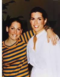
(Susan Komen on the left; sister Nancy Brinker on the right)
Susan grew up to be the “darling of Peoria,” a beauty queen and local model. She would enter a disastrous first marriage (groom collapsed at the wedding for starters), followed by a second marriage to Stan Komen who would run a liquor store – Stan’s Wine and Spirits – in Peoria until his retirement in 2014.
At age 33, Susan felt a breast lump. Not a good thing in 1977, on the eve of a revolution about to occur in the management of breast cancer. 1977 was, however, the peak time of enthusiasm for “subcutaneous mastectomies” with the newfangled breast implants for reconstruction…sometimes patient-driven, sometimes surgeon-driven and sometimes both. Pain, cysts, “fibrocystic disease,” you name it, then cough up the money, and surgeons would perform the “scoop out” procedure with implant reconstruction. Some women were very happy with their result. Many were not. And for some, considerable amounts of breast tissue remained in place. Nevertheless, no one at the time conceived of using the procedure for treating cancer — except for Susan’s surgeon who had been recommended by her family physician in Peoria.
After performing subcutaneous mastectomy for Susan’s cancer, the surgeon pronounced her cancer-free, a tad premature since she developed positive nodes shortly thereafter and systemic metastases as well. After treatment at both the Mayo Clinic and M. D. Anderson, she was still left with chest wall recurrences and died 3 years later in 1980 at the age of 36. Given the aggressiveness of this tumor, it is hard to lay blame on the unusual subcutaneous approach used for local control, but it is noteworthy nonetheless.
Meanwhile, younger sister Nancy had moved to Dallas where she became a buyer for Neiman Marcus, and then, in 1981, she became the bride of Norman Brinker, the restaurant magnate who founded Steak and Ale, et al, (now Brinker International) and is credited with the development of that omnipresent dining staple – the salad bar. With her new financial stature and a deathbed promise to her sister Susan, Nancy Brinker founded the Susan G. Komen Breast Cancer Foundation in 1982, a mere 2 years after Susan’s death. The foundation became intimately linked to Dallas as a result of Nancy’s new home, not Susan’s home in Peoria (although Peoria did become a site for a “Komen Breast Center,” a nationwide network concept of screening centers that never happened.)
The first Race for the Cure took place in Dallas, Texas with 800 participants in 1983. The rest is history. I attended the first race in Oklahoma City (1989, as I recall) where participants raced around the track at Remington Park. Pink ribbons were not introduced until 1991, and though several groups lay claim to the pink revolution, I’m pretty sure it was a Komen innovation.
One year after that first Race for the Cure, Nancy Brinker was diagnosed with breast cancer. She underwent unilateral mastectomy and later contralateral prevention, even though her BRCA-1 mutation was not confirmed until 2006.
Initially, the sole agenda for Komen was to flood the country with high quality screening mammography. This drew some objections, including one prominent breast surgeon, the late Susan Love, who broke ranks with the movement by clarifying that mammography is not a “cure,” and that we needed to be thinking more about a true cure and a “post-mammographic era.” Eventually, Komen expanded its scope to all types of breast cancer research.
Nevertheless, the growth of the Race for the Cure was phenomenal, taking place in a parallel fashion to the breast cancer diagnosis and treatment revolution that was well underway. It is estimated that, today, over 1.5 million participants raise money through one of several outreach programs sponsored by Susan G. Komen (several name changes of the foundation have occurred over the years, but Susan’s name is always there). Politics, of course, wormed its way into the activities of Susan G. Komen and eventually Nancy Brinker resigned as CEO.
Back to Breast Cancer Awareness in the spooky month of October. Around 1985 or 1986, the American Cancer Society teamed up with a pharmaceutical company that later became Zeneca, then AstraZeneca, announcing October as National Breast Cancer Awareness month. Zeneca was criticized for self-serving interest since they manufactured pharmaceuticals used for breast cancer. But they had, in fact, done their own internal audit on employees, showing that it was cheaper to screen with mammograms for an early diagnosis than to do nothing and pay for treatment of more advanced disease.
At this same time, the famous (or infamous) Breast Cancer Detection Demonstration Project was reporting results indicating that massive screening of the general population in the U.S. was both feasible and effective. The BCDDP was sponsored by the American Cancer Society and the National Cancer Institute, both organizations riding the waves of the War on Cancer legislation signed by President Nixon in 1971. So, by the mid-80s, the policy makers were wildly enthusiastic about general population screening with mammography, and it was a case of “full steam ahead.”
Still…why October?
I’ve not been able to nail down the exact details surrounding October as Breast Cancer Awareness Month, but I have a theory — a link between the rapid rise of the Susan G. Komen Foundation and the proclamation about October made by the American Cancer Society. I think the answer might be found by looking at Susan Goodman Komen’s birthday – she was born on October 31, 1943. She would have been 81 this Halloween.
In 1976, pioneering breast radiologist, John Wolfe, published results he’d been working on for the prior decade — that is, certain mammographic patterns being associated with an increased risk for breast cancer. This wasn’t your measly “4-6X” risk that emerged (with much confusion) in modern times, but a 37-fold increased risk imparted by the now-infamous “DY” pattern. Translated to absolute risk, that’s 45% breast cancer risk for women aged 40-59, and a whopping 82% risk if focused only on the 50-59 age group. And with that in mind, Dr. Wolfe recommended that those women identified with the DY pattern undergo preventive subcutaneous mastectomies, a new procedure being promoted by the plastic surgeons (for a variety of indications) upon the introduction of silicone implants.
The curious thing about Wolfe’s landmark article and his recommendations is that he divided mammographic patterns into 4 categories (the same number we have today), but the divisions were based on breast cancer risk, NOT the likelihood of missed cancer, i.e., “sensitivity.” The N1 category was “predominantly fatty” and was associated with such a low cancer risk that screening was not needed. P1 meant less than 25% “prominent ducts” and again, low risk for cancer, so no pressing reason to screen. P2 was defined as greater than 25% “prominent ducts” and screening was now advised due to the risk status. DY was dense fibroglandular tissue with very high risk (analogous to today’s Level D), to the point that Wolfe made his remarkable recommendation for preventive surgeries for all DYs. In 1980, when I began private practice in Los Angeles, all mammography reports included the Wolfe classification, though we had no idea what purpose it served since interventions were unproven, and screening recommendations were new. None of the recommendations from various organizations included Wolfe in their guidelines.
Turns out, Wolfe’s methodology was flawed for a variety of reasons. First of all, most of the women he studied had symptoms of some sort. That is, they were not asymptomatic, which is required today before calling a study “screening.” So, for example, when a patient in his study with a DY pattern developed breast cancer 7 months after a negative mammogram, Wolfe felt the DY pattern, as a strong risk factor, had accurately predicted a future cancer. But this wasn’t the case at all! He was witnessing the extraordinarily high “miss rate” for mammography when women have dense breast tissue. The cancer was there on the prior mammogram, but not visible due to the camouflaging effect of density! He was correctly observing a relationship between density levels and breast cancer, but misunderstanding the true reason for his observations. Ever since then, we have struggled to recognize that there are two related, albeit independent, implications of high mammographic density — 1) a modest risk factor, and 2) the strongest predictor for missing a cancer on routine screening (“poor sensitivity”).”Wolfe patterns” came and went, and after that, trying to relate breast cancer risk to mammographic patterns fell into a state of hibernation. Any attempts to document increased risk associated with density patterns went unnoticed by clinicians. We had moved “beyond Wolfe,” it seemed. At the same time, an enormous effort was engineered by the American Cancer Society and others to promote mammographic screening. In these efforts, mammographic sensitivity was said to be “90-95%.” As it turns out, this “90-95%” was based on a large feasibility study (the BCDDP, not the randomized controlled trials that proved screening saves lives) where many of those entering the massive BCDDP study had palpable masses. Stage II cancers outnumbered Stage 0 and Stage I combined. “90-95%” was never true for asymptomatic women, yet it dominated thought for another decade or so. Few clinicians were paying attention to the rather large number of cancers that were being missed on screening mammograms. This should be no surprise. How do you recognize and count the number of missed cancers if you’ve missed them already? The standard approach was to count cancers that emerged after negative mammograms for a period of 12 months of follow-up, but this was arbitrary and unreliable. (Adjunct imaging to identify missed cancers was not yet available.) For palpable cancers — yes, the sensitivity was 90%. But that’s not screening, and we failed to make that distinction in those early years. The definition of mammographic screening evolved to imply asymptomatic patients (no palpable masses), and it was here that the sensitivity of mammography was largely unknown to clinicians.
Then, technical developments took place in the 1990s with ultrasound (US). No longer was US just a matter of cyst vs. solid, but it could distinguish benign vs. malignant. And with this development, many radiologists launched studies to screen asymptomatic women with US, after negative mammograms…IF the breast tissue was dense. There were no definitive trials at first, that is, no prospective randomized studies. But the results with US were numerically consistent across the board. Mammograms weren’t coming close to 90-95% sensitivity in women with dense breasts. Completely independent of “future risk,” breast density was predicting mammographic failure due to the camouflage effect. Arguably, this failure to detect cancer is much more significant than the modest risk imparted by density (more on this below).
Then, in the early 2000s, the introduction of MRI into the breast clinic turned the sensitivity problem upside down — that is, mammography alone in high-risk women was missing more cancers than it was finding. Sensitivity was 40% across the board in the MRI screening trials. This was a far cry from the “90-95%” starting point 25-30 years earlier. Granted, MRI lowered the threshold of detection to smaller tumors, which made both mammography and US look worse. On the other hand, ultrasound did not lower the threshold of detection, so for purists, US probably reflects the miss rate of mammography more fairly. Nevertheless, what MRI is doing becomes clear when the numbers are examined — MRI is finding current breast cancers that are invisible on mammography and/or US, then — by lowering the threshold of detection, it is also finding most of the breast cancers that won’t be discovered until the next routine screening study, 1 or 2 years away.
Back to Breast Density. Overcoming the “false alarm” by Wolfe was not easy. I was once part of a team of primarily academic radiologists who taught MRI to other radiologists around the country, shortly after MRI was introduced. I was the token surgeon. In one of my talks on MRI screening guidelines, I pointed out the overwhelming evidence that breast density was an independent risk factor for breast cancer development. This was nowhere near the Wolfe levels, but the positive results were similar in more than 10 studies. So, why was no one paying attention? I don’t know. Were they remembering the collapse of the Wolfe criteria? In my presentations, I likened breast density to comedian Rodney Dangerfield — “No Respect.” Breast density was not part of the mathematical models that estimate risk. It was not used clinically with regard to any screening recommendations with any modality (It’s still not considered a problem requiring multi-modality imaging by the U.S. Preventive Services Task Force!). And, when added to the MAJOR problem of failing to detect breast cancer, density was — well — it was Rodney Dangerfield’s calling card: “I don’t get no respect.”
I don’t think I made a dent in the lack of interest in breast density. What made the difference was a grassroots movement launched when women discovered the truth – that many of them had mammograms so dense that cancer detection was nearly impossible, yet no one told them! (I’ve covered this story about the late Nancy Cappello in other blogatorials.) Today, we celebrate legislation that forces radiologists to talk to patients about this problem, but how in the world did we get to the point where this had to be legislated? I started a High Density counseling program and clinic at my facility just as soon as we opened our MRI screening program in 2003. And, I published a point system for MRI screening where Density played an equal role to Risk level. In my mind, density was even more important than risk level. After all, why talk about a second form of imaging if you don’t take into account the failure rate of the first modality?
Yet, when the American Cancer Society released their 2007 guidelines for MRI screening, “Density” was relegated to the “need more research” category. And it’s been a brick wall ever since. We also published our local MRI screening results before and after the ACS guidelines, wherein one-half of our MRI-discovered cancers would not have been found if we’d followed the ACS guidelines. By including density, our MRI cancer yield doubled. Europe is way ahead of the U.S. on this one. The European Society of Breast Imaging has recently added a screening approach for women with Level D density, with no other risk factors: “Breast MRI every 2-4 years.”
So, even though radiologists must divulge high density levels to patients, not everyone will counsel patients at that point as to options. Even if options are discussed, the breast center must understand that insurance often will not pay for high-density screening beyond mammography (following the Task Force guidelines). So, the breast center should have low-cost programs for cash customers who have opted for US or MRI (but that’s another story — many hospital-based breast centers don’t like that “low-cost option.”)
Now, in spite of the powerful presence of “breast density” in the screening world today, there are points of confusion still circulating. I’ll mention 6 of them:
“Density imparts a 4-6 fold risk of breast cancer.” This is highly misleading, as this is a “relative risk,” and all relative risks have a numerator and a denominator. In this case, the denominator is those women with fatty replacement pattern (Level A). This is only 10% of the population and hardly “average.” So, yes, comparing Level D to Level A generates “4-6 fold risk.” But if you compare Level D to the “average patient” then the Relative Risk becomes a much more manageable 2-fold risk. This misconception was so pervasive initially that huge efforts were made to educate physicians about these numbers, and the problem has been largely corrected. Most clinicians have been exposed now to this misconception, though it still leaks out occasionally.
“The risk imparted by density is now covered nicely by the Tyrer-Cuzick (TC) model.” Careful. The referent (where RR=1.0) in the TC model is essentially Level C density. Not Level A. Not Level B. So now, patients who have been labeled as “dense” Level C and a slight increase in breast cancer risk will actually have Zero risk (RR=1.0) added by density using Version 8.0 of the TC model. So, “high risk” when speaking of dense breasts in general, but “no elevated risk” for Level C when using the TC model. Only Level D translates to higher absolute risk in the TC model. Level C is neutral, while Levels A & B will actually generate lower levels of risk with the newer Version 8.0 (RRs less than 1.0).
“Dense and non-dense is the currently preferred approachin defining women for specialized screening.” What we really have is a density continuum from 0 to 100% (the practice of medicine “hates” continuums, as we need to stratify our strategies.) Furthermore, we have a classic Bell Curve, with most patients bunched up in the middle where the Level B and Level C patients dominate, accounting for 80% of the total. So, when we turn this into a dichotomy, we draw a line down the middle of the Bell Curve, and there is little difference between a high Level B and a low Level C. In fact, it is highly subjective, and radiologists don’t necessarily agree; even a single radiologist can be inconsistent. Yet, this is where most women are due to the Bell curve — either “high B or low C.” But the “dense and non-dense” dichotomy (A/B or C/D) implies a sharp distinction that’s just not there.
“Software is now available to take away the subjectivity.” While it is impressive to see exact percentages emerge with these tools, you can’t get away from the Bell Curve. Most women are clustered right at the division line, with huge differences in management depending on which side one falls. This limitation (confusion?) has been created through the simultaneous use of a dichotomy (by the grassroots movement) and the 4 Levels A-D from the American College of Radiology. If we’re going to use 4 levels, then each level should have unique recommendations. Another problem that is not addressed by software packages — there is a qualitative feature of density in addition to the well-known quantitative. This qualitative aspect is exceedingly complex and beyond discussion here. However, to give a practical example, I create a dime-sized mass in my head, then move it throughout the white areas of a mammogram to see if there’s somewhere a cancer could hide, even in low density patients. If you are a clinician, think about those mammograms where the only density is a homogeneous white sheet confined to the upper outer quadrants. This will be a Level B patient with only 25% density, but if cancer develops in the UOQ, it’s going to hidden, just as if the patient were Level D. Remember, overall breast density is merely a surrogate for the density that surrounds a cancer. And, this could even be a worse problem, considering that these dense patches might be where cancer originates. (P.S. If you think this sounds overly compulsive (nuts), then read about the Entry requirements for ACRIN 6666 (density + one additional risk factor, studying multimodality screening). A patient could qualify with a low overall density if there was a homogeneous white region involving just one quadrant in each breast. Level A in 3 quadrants, Level D in one quadrant, but instead of a fusion to Level B, patients qualified for ACRIN 6666). You can see that the risk of missing a cancer on mammography has this qualitative aspect in addition to the 4 quantified levels.
“Density disappears with Age.” Radiologists know this isn’t true, but many clinicians (and epidemiologists who set screening guidelines) believe that extra imaging with US or MRI will not be needed after a certain age (60, for example) because low density translates to mammographic accuracy. While this transition to less density might be true overall in a large cohort, there are many exceptions to this rule. We’ve all seen the 70 y/o with Level D density. And, when encountering the young woman with Level D density, it might improve somewhat over time, but she is going to remain dense her whole life, even though a shift down is possible. This controversy comes up in the MRI screening guidelines where older women are discriminated against with “lifetime risk” calculations. Older women have passed through much of their lifetime risk, so they don’t qualify for MRI screening, yet their short-term risk might be very high — much higher than a younger woman who qualifies on the basis of many years left in her lifetime risk. I’ve tried to make this point in publications and at screening guideline meetings, and am met with, “the MRI guidelines were intentionally designed for younger women with dense breasts.” Fine. Agreed. But what about the older woman with dense breasts? They can be included without taking away any of the benefit for younger women. And, the answer is so easy — just add short-term risk calculations alongside the lifetime calculations. On this point, I’ve made about as much headway as Rodney Dangerfield.
“3D tomosynthesis has solved the density problem.” Or, the way a patient might state the issue — I’d like to stop Ultrasound (or MRI) now that we have 3D mammography.” Sorry, but 3D is not even close to solving the density problem. It helps, yes, but let me put this in perspective. Shortly after the introduction of 3D, an Italian study pitted 3D mammography compared to old-fashioned 2D plus Ultrasound. It wasn’t even close. The combination of 2D plus US discovered many more cancers than 3D. It gets worse. A clinical trial in the U.S. pitted breast MRI against 3D mammography. MRI found 95.7% of the cancers while 3D found…wait for it….39.1%.
Is there any hope for the patient with dense breasts, short of MRI? Yes. While Ultrasound is currently thought to be the next best thing to MRI in high-density screening, Contrast-enhanced mammography (CEM) is more sensitive than ultrasound and might be the answer. We’ve already seen the numbers. CEM is using a radiologic dye, injected prior to the 3D mammogram. Results appear to be in the same ballpark with MRI, though more definitive studies are being performed now. We know enough already that CEM can be recommended when patients are unable to undergo MRI for some reason. And, CEM is easier on patients, and cheaper. The question is whether or not it can replace MRI rather than its current role as a comparable back-up.
PS – I’d be remiss if I didn’t mention the ultimate answer (in my mind) to all the mess above, and even more mess I haven’t addressed with using Risks and Density to guide adjunct imaging recommendations. If we had a reliable screening blood test, then women could undergo mammography and blood testing as a matter of routine. And if mammograms were negative, but blood test positive, this would be the signal for adjunct US, CEM, or MRI. (That’s why I’ve been pursuing this agenda for the past 30 years.)
I’m going to end it there, but with a plug for a new breast center opening soon in OKC (SW 89th & I-44), where we will have Contrast Enhanced Mammography as an option, as well as a High-Density clinic to accompany the High-Risk Clinic. More information to follow, but the Premier Breast Health Institute of Oklahoma will be opening its doors in the fall, 2023.
Over the course of 25 years, I watched The (Daily) Oklahoman expand its Book Section from a one-pager once a week to a 2-pager where Oklahoma authors and their long-suffering efforts could be featured. And, there was a point person, that is, a book editor, who freely communicated with authors. Then it all imploded. With USA Today taking over The Oklahoman, many local authors returned to being second-class citizens. Currently, syndicated reviews occupy less than a half-page, and for local authors, the great hope is that, in the absence of a formal review, one’s upcoming signings will be listed (my last signing didn’t even make it that far). Regardless, I originally thought I might be able to sneak in a review of my trilogy – The Brainbow Chronicles – or, at least the first book — the award-winning NUTSHELL. My intent in the query email was to humbly brag that, 23 years ago, I had a successful novel reviewed in USA Today (not the local rag, but the Big Kahuna), and that I was merely inquiring about a review to appear in the OKC version of the newspaper. Slam dunk. Right?
After I sent a copy of the book to the newspaper to accompany the email, the response was nil. Nothing. Silence. Zip. Not even a thank-you for the book. Later, it was the same response after mailing the subsequent books as they were released over a 2-year period. Next, I figured I would bypass the local staff, especially since the OKC newspaper doesn’t have anyone designated as Book Editor. I sent my next query directly to the Big Kahuna USA Today offices (where they DO have Book Editors) since I’m in the club (sorta) of authors who have made USA Today national headlines with their books (FLATBELLIES in this case). On the bright side, they did not reject the offer; then again, they didn’t accept the offer. You guessed it. No response at all.
I understand the New Ethics don’t require you to answer anyone you don’t feel like answering, but it’s still amazing given the ease of email. In the days when writing a letter was comparatively tedious, a query in the book business was always answered, even if it generated only a form letter. But today, the mantra is: “I don’t have the 5 seconds it takes to write ‘no thanks’ and press SEND.” You might call this “ghosting,” but you would be wrong on a technicality — “ghosting” requires a relationship prior to cutting off all forms of communication. My ghost was never alive.
Marketing one’s book through self-promotion is awkward, embarrassing, tedious for friends and family who serve as beta readers, and simply no fun. And if you have a small publisher, you are going to spend 10X the effort for one-tenth the success. (When the Big Kahuna reviewed FLATBELLIES, I didn’t lift a finger. The publisher arranged it all.) So, faced with the New Ethics, I figured it’s better to return to one’s hometown, where the venerable newspaper still functions by the Old Ethics, in this case, the El Reno Tribune, with editor Ray Dyer. Having followed my writing career, Mr. Dyer suggested a feature article about my life as an author rather than the standard book review. Since my writing history pre-dates FLATBELLIES, we ended up with a long article that spills off the front page to include 2 more pages (links below). Of course, there is fairly strong interest in El Reno, given that my most successful book, FLATBELLIES, was based in a town called “El Viento,” and it’s no secret that this coming-of-age novel is semi-autobiographical.
In the article, you might be surprised to learn that my first book was NOT Flatbellies. In fact, I wrote a book in 1977 that had greater overall potential than Flatbellies. The “story of this story” is surrealistic even today. Here’s a teaser: As a surgery resident-in-training, I walked out of the VA hospital in OKC on a Saturday, flew to Los Angeles to sign my “rich and famous” contract with a prominent Hollywood agent, then flew back to OKC in time to take call on Sunday. I hope to tell the complete story someday, perhaps along with the never-published PROGNOSIS: GUARDED. When it all came crashing down, extinguishing the stars in my eyes, it was FLATBELLIES that emerged from the rubble, over 20 years later.
If you read the article in 3 parts below, be aware that you’ll probably need to Zoom at least 100% to read clearly. And to ZOOM, you might have to access the controls listed at the bottom as “pdf tools.”
Thanks again to Editor Ray Dyer of the El Reno Tribune.
Here are the links in 3 parts, top to bottom, to access the article in the El Reno Tribune:
Until 2009, few clinicians had heard of the U.S. Preventive Services Task Force (USPSTF), but in fact, they’ve been around since 1989 when they first endorsed screening mammograms, starting at age 40. The controversy about the “starting age” actually began in the late 1960s, and was already a hot topic for debate in the 1970s. Professional organizations were split down the middle, half favoring 40 and the other half favoring 50 as the starting age (supposedly based on data, not emotion)
The Task Force was conceived as an independent group of experts in public health and screening programs. Today, 16 members monitor over 100 screening recommendations for all sorts of diseases, with updates every few years. The Task Force is quick to point out that they are not a governmental agency, but they are funded by the government, with oversight by the government, so what does it matter? To complicate things, members rotate through the committee with relatively short terms, which might explain their remarkable inconsistency with regard to breast cancer screening. For breast screening recommendations, the USPSTF tries to update their position every 6-7 years, based on “new evidence.” Yet, if you read closely, that “new evidence” is often related only to the harms of screening (such as psychological trauma with a benign biopsy). Meanwhile, the benefits are calculated using obsolete data from primitive mammography used in the historic trials of the 1970s and 1980s. “Well, it’s all we’ve got,” they announce pitifully, while ignoring a mountain of data that supports screening average risk women beginning at age 40.
And while few had heard of the USPSTF in 2002, everyone heard by 2009 when they shocked the breast cancer community (and the world) with “Start mammograms at age 50, then every 2 years.” Those who understood the impact quickly calculated how many women would die unnecessarily each year due to these recommendations (“many thousands” for purposes here). So, by following the rules of “good science” and “evidence-based medicine” here’s what the USPSTF has done to make fools of themselves with regard to screening at 40 vs. 50:
1989 — start at 40
1996 — no policy, 40 or 50 is fine
2002 — start at 40
2009 — start at 50 and screen every 2 years
2016 — same as 2009
2023 — start at 40 and screen every 2 years
Meanwhile, other organizations (including the American Cancer Society) said “start at 40” throughout the entire controversy. Only recently, did the ACS switch to 45 to try and mediate the bitter arguments generated by the USPSTF. Of course, the American College of Radiology has maintained “start at 40” throughout the decades, but sad to say, no one trusts experts anymore. They are allegedly biased for their own personal gain, so insurers pay no attention to them. But what about “mostly neutral” organizations like the American Society of Breast Surgeons who make no money through screening programs? Doesn’t matter. They’re “friends” of the radiologists, and they can’t be trusted either. (Point of information and disclosure: I was a member of the most recent Screening Guideline Committee of the American Society of Breast Surgeons, and our group was nearly unanimous — “start at 40 with annual mammograms”).
The American Academy of Family Physicians, however, strongly (blindly?) endorses the USPSTF, as does the American College of Physicians (internal medicine specialists), making it so hard to battle the USPSTF that my own hospital in 2009 began telling its employees to wait until age 50 to begin screening (while I’m serving at the national level, endorsing 40). And I can assure you my hospital didn’t give a rip about what I said, or what Dr. Stough said, because the employee guidelines were drawn up by members of the American College of Physicians (read: USPSTF), located at the St. Louis hub, who voted along party lines — hook, line, and sinker — for age 50.
Screening is far more complicated than most imagine. I became heavily involved as an advocate of MRI screening, which was tied to risk assessment and genetic testing. I even started writing a book on MRI screening, getting about halfway in my efforts when the 2009 tsunami hit, and we went from 40 to 50. I abandoned my MRI intent, seeing a greater need to clarify screening in general, including the history of breast cancer screening, and how deeply flawed the USPSTF was in their assessment. When I finished, my publisher asked me to provide 10 possible titles for the book. They rejected all 10, not wanting to see “MRI” in the title. They renamed my book, “Mammography and Early Breast Cancer Detection: how screening saves lives.” Even today, I don’t know of any book that covers the controversy like I did. Sadly, few have even heard of the book, but I’ve received personal comments from experts around the world. One retired academic radiologist from the University of Manchester, U.K. wrote this: “I wish I had this book back when I was training residents in breast imaging. Everyone doing breast imaging should read it. To be honest, even I didn’t fully understand the controversies and complexities surrounding screening.”
There’s a huge difference between the skills needed to interpret mammograms and the epidemiology behind general population screening. Most practicing radiologists are unaware of the theories of screening and how statistics can be twisted. They might be superb at identifying cancer on a mammogram, but that has nothing to do with a controversy like “overdiagnosis.” But by the same token, the epidemiologists and others of the Task Force ilk do not understand breast radiology, leaving them often with empty-headed opinions.
Yes, you can use high-powered stats and sophisticated means of twisting those statistics to assess both the BENEFITS of screening and the HARMS of screening. But after the necessary calculations are made, and you put the two on a scale…they cannot be balanced! It’s entirely subjective at this point. BENEFITS vs. RISKS. How many benign biopsies does it take to neutralize the benefit of an early detection? It’s the final balancing act that keeps this controversy from being purely scientific.
I learned about the 2023 Task Force update like most everyone else — watching the evening news, then online coverage. Sounded simple, and as the USPSTF committee member told us about the NEW guidelines, it was crocodile tears as far as I was concerned (“We must be attentive to the fact that breast cancer incidence is increasing in younger women.” But who abandoned those women in 2009?).
Another neglected issue: younger women often need more than mammography, i.e., ultrasound at a minimum and perhaps MRI. And yet, the Task Force has been irresponsible in ignoring the data we have now on ultrasound screening and MRI screening. Guess what the Task Force says about breast density….”no proof that there’s any benefit from adding ANYTHING to mammography in women with dense breasts — that means no 3D, no ultrasound, no MRI, no molecular imaging, and no to anything else you can think of.” That leaves digital mammography as the sole recommendation, yet of all the types of imaging I just listed, 2D digital mammograms are the WORST at detecting cancer, or scientifically speaking, 2D mammograms have the lowest sensitivity for cancer detection among the wide array of options. And THAT’S the recommendation of the Task Force. I consider their approach to dense breasts to be an act of “malpractice by committee.” And if you think that’s harsh, there are applicable words that make “malpractice” sound soft. The Task Force has done more to harm breast cancer screening than any other organization — even more than those mammography-deniers who don’t believe mammograms save ANY lives. The difference between the Deniers and the Task Force? The Task Force claims, “We’re the authority on this, and we’re here to help you.”
The emergence and adoption of breast cancer screening is a remarkable story of the power of human bias. So, rather than the MRI book I had envisioned, it ended up being a book about screening for breast cancer in general, a soap opera when it comes to the history that continues to this day. Although the genre of the book is classified as “medical text,” it’s anything but. It’s human drama, the same as my novels. Sadly, the book got stuck with a bad title and a bad cover. But as one reviewer said, “In this era of doing less screening, Hollingsworth makes a convincing case that we ought to be doing more, not less. And for sheer entertainment, don’t miss the Endnotes.” (In fact, I had placed what became endnotes in the text itself, spicing up the story. But the editor supervened again, and pulled out many of the juicy tidbits and placed them in the Endnotes).
As I review my Task Force critiques throughout the years, I find THREE Powerpoint talks that I used to give to physicians at CME courses — one talk is 30 minutes, one is 45 minutes, and the one I preferred to give lasted a complete 60 minutes. Then, most of the book I’ve already mentioned is tied to the same topic. And finally, many of the essays in my book The Best Breast Blogatorials cover the Task Force and their wily ways. So, to make sure crocodile tears on the evening news don’t sway you, read one or both of my breast cancer books.
PS: I’m having trouble providing links in this Blog template, so the Blogatorial book will need a search using the title/author.
On March 10, 2023, the FDA issued a rule making it a requirement to send letters to patients undergoing screening mammography to inform those who have dense breasts. Where do I begin? After all, my professional study of mammograms that miss cancers dates back 30 years with my first academic paper: “Establishing a Histologic Basis for False-negative Mammograms,” published in the American Journal of Surgery….and the only publication of mine that was ever honored later as “One of the Top Contributions to Oncology in the Past 50 Years” by the same journal in a later anniversary issue.
Funny thing, though. At that time, I was focused on the impact of histology of various types of breast cancer, that is, what tumors look like under the microscope. And it was clear that some tumor growth patterns escape detection by mammography because they grow diffusely, without forming a “mass.” That publication was only the second time that “invasive lobular” cancer had been implicated in false-negative mammograms (common knowledge today). And, we made brief reference to the additional theory that tumors might be missed on mammography due to breast density. Only a few authors had raised this question….in 1993.
Long story short, in writing that first article, I wanted to start off with what was known about mammographic sensitivity, that is, the percentage of cancers detected. To my dismay, I could not find the reference that supported the oft-quoted, official, widely accepted… 90% sensitivity. I’ve chronicled this search in my books, and more recently, as the most comprehensive coverage of the story, written in a review article published in the American Journal of Surgery (same journal as above, circling back from my first publication). It’s a fascinating story of scientific paralysis, where the medical community refused to accept the true miss-rate of screening mammograms, a problem that continues to this day.
The question of missed cancers was hard to answer back then: How do you know you missed a cancer if it’s not on the mammogram? Quick Answer: you don’t. Instead, they used arbitrary follow-up periods, usually one year — any cancer appearing quicker than one year after a negative mammogram was a “miss.” It would take many pages to explain why this was an absolutely terrible way of judging sensitivity. But when breast ultrasound was introduced, the cracks in 90% started to form, and by the time breast MRI was introduced to the clinic in 2002, the belief in 90% was shattered (for many, but not all). If MRI is compared to mammography head-to-head, then we discover the chilling truth that mammograms miss 60% of cancers. That’s right, the number is remarkably similar from study to study. The sensitivity of mammography is only 40% when compared to MRI….and that includes all levels of density. If we restrict this to Level D density (extreme), then mammographic sensitivity for these women is around 20%. Stated alternatively, if mammograms tried to get FDA approval today, they would be rejected for Level D women and perhaps Level C as well.
When breast MRI first came on the scene, the “father of breast MRI,” Dr. Steven Harms, asked me to join his team of radiologists who educated the rest of the country’s radiologists on the use and interpretation of breast MRI. We traveled to so many cities I can’t even recall where. Nevertheless, one of my jobs as the token surgeon was to “crack the 90%”…”Do that 90% thing you do about mammography” wherein I would share the bizarre story of how we ended up with a grossly exaggerated number that doctors and patients grabbed onto, then refused to let go.
Back to the FDA. What are they doing sticking their collective noses into how we report screening mammography results? Well, little known fact — breast imaging became the first radiologic test governed by the FDA in the early 1980s. Sad to say, when mammograms were introduced in the 1970s, “mammography factories” opened up, and did a terrible job of quality assurance. Horror stories were often exposed on the evening news. The FDA took over, and still to this day, every breast center has to be FDA approved, then undergo a yearly inspection.
Here’s where it gets weird. For years, through state legislation (Oklahoma passed its law about 5 years ago), nearly every state in the Union already had this legislation in place (hard to believe it required legislation of any type, isn’t it?) So, the FDA has made it 100% (all states) and has standardized the reporting. So what? The final few states now have to comply, but that’s about it. There are no guidelines for these women who are left floundering, and are not counseled accordingly. It’s not an easy subject, and I won’t dwell on the mountains of misinformation that are out there for women with dense breasts. Oh, yes, the American College of Radiology issued guidelines, recommending the addition of breast ultrasound to mammography for women with Level C and D mammography, and this is all well and good. Problem is, we live in an upside-down world where the expert providers of medical service are not allowed to establish their own guidelines because of “known bias.” So, neutral organizations have to take the leap and confirm the need for additional imaging in women with dense breasts because third-party payors won’t pay attention to the expert clinicians.
Now, let me take it to a different plane. A few states (e.g., Connecticut) have lobbied for insurance coverage for screening US added to mammography for women with dense breasts and have WON. Great. But compliance is a huge disappointment (they might be up to 25% by now). Everyone drank the Kool-Aid of 90% sensitivity for mammos years ago, and now, even when covered by insurance, women settle for mammograms alone, and ignore those who are shouting: “Mammograms are not enough!”
This is why, btw, that I continue blood test research (currently with Syantra, Inc) so that women who have settled for mammograms alone will be inspired to proceed with additional imaging if the blood test is positive.
With my research career still active, we are ramping up for a definitive clinical trial of SyantraDX in high-risk and/or high-density women with normal mammograms who would then undergo additional imaging if blood-test-positive.
My coverage of the true complexity of the breast density problem and the fascinating history of the 90% can be found elsewhere on this web site, or in either of my two breast cancer books. It’s a fascinating story, especially the breakthrough in awareness that began with a grassroots movement prompted by one woman whose mammograms failed her miserably. Read about it here:
Mammography and Early Breast Cancer Detction: how screening saves lives
The Best Breast Blogatorials (essays taken from this web site)
I continue to fight the fight. MRI breast cancer screening guidelines were flawed from the beginning in 2007, and I’ve tried my best to draw attention to major issues that need to be corrected. I even served on the Screening Guideline Committee for the American Society of Breast Surgeons, and still couldn’t conjure any major changes. My major point is that breast density barely fits into screening algorithms, in spite of all the attention it gets today. But if you’re proposing a two-method imaging strategy, shouldn’t the first question be: “What is the probability that mammograms will fail?” That probability is based on density…yet the question is never asked using current guidelines. Instead, density is relegated to a modest risk factor (which is unrelated to whether or not a cancer will be found on MRI).
Evidence that clinicians are coming out of hibernation on this issue has finally arrived — The DENSE trial in The Netherlands is now reporting results greatly favoring MRI over mammography based on a single criterion — breast density. If you have Level D mammograms, you qualify. Very simple. And from that trial, the European Society of Breast Imaging, in 2023, changed MRI screening guidelines to include women with no other risk factors than Level D mammograms, to be done every 2-4 years in addition to mammography.
This idea of extending the intervals for MRI for women at lower risk levels (although density is a modest risk in and of itself) was first proposed in 2008 where a point system gave equal weight to density and to risk. In this system, the points are added, and the total number dictates the interval for MRI — every 1, 2, 3 years or not at all. This point system was published in Seminars in Breast Disease (now defunct) by my radiologist colleague, Dr. Rebecca Stough and me in 2008, a full 15 years before these new MRI screening guidelines with longer intervals from Europe.
At the time, we had our initial experience with MRI to go on, but after that, it was common sense, often dismissed as “weak” rational thought in this era of evidence-based medicine. That said, our system that focused on density yielded DOUBLE the number of MRI-detected cancers as the American Cancer Society’s guidelines. Those 2007 guidelines from the ACS remain the same. Oher organizations in the U.S. have made modifications to the original, but have not corrected most of the issues we have brought to light in a series of critical editorials.
A few weeks ago, we had an Editorial published in the online journal CUREUS, calling for a complete overhaul of the MRI screening guidelines in order to allow density to play a greater role.
Here’s the link to the new article about breast density and MRI guidelines: bit.ly/3Mh22uz
ABH
PS: I wonder if there has ever been simultaneous publication of a medical article and a medical novel at the same time? If you’re more interested in my novels, esp the new trilogy by “John Albedo, here’s the link to the book trailer: https://bit.ly/3lzaRot